Deep learning: Improving materials characterization through automated EELS analysis

Deep learning is a promising tool for the automated characterization of materials at the nanoscale using electron energy loss spectroscopy.
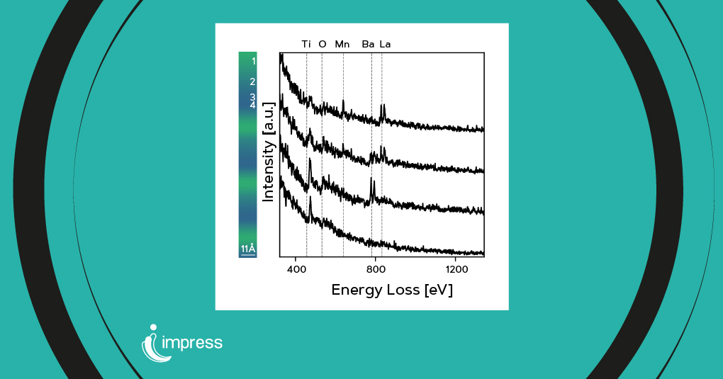
Deep learning is increasingly recognized as a powerful tool for advancing materials characterization, particularly in Electron Energy Loss Spectroscopy (EELS), as highlighted by a recent study. Traditionally, identifying core-loss edges and their corresponding elements in EELS spectra has required manual effort, making the process time-consuming and error-prone, especially in large datasets.
The potential of deep learning in electron microscopy
To address these challenges, a team of researchers from the University of Antwerp in Belgium, one of the IMPRESS project partners, have developed a synthetic dataset to train various neural network architectures, including convolutional neural networks and vision transformers. The ensemble of neural networks achieved remarkable performance, enabling fully automated elemental mapping in EELS spectrum images.
Automated materials characterization
The results of this innovative study were published in Scientific Reports, a journal within the Nature Portfolio. The study focused in particular on EELS, a technique that provides detailed elemental information at the nanoscale. Johan Verbeeck and colleagues developed a synthetic dataset comprising 736,000 labeled EELS spectra, representing 107 core-loss edges and 80 chemical elements to train a series of neural network architectures. The scientists subsequently used an ensemble of neural networks to further increase performance.
This approach demonstrated the effectiveness of fully automated elemental mapping in a spectrum image by directly mapping the predicted elemental content and using the predicted content as input for physical model-based mapping.
A leap forward in electron microscopy techniques
These findings highlight the potential of deep learning in electron microscopy, aligning with the IMPRESS project’s objectives to integrate innovative developments with a novel interoperable platform for transmission electron microscopy. Thanks to these advancements, electron microscopy techniques will be improved and made available to a broader range of users, thus enhancing nanoscale research capabilities.
“Our study exemplifies how innovative tools, such as deep learning, can improve materials characterization and be seamlessly integrated into advanced electron microscopy platforms, making nanoscale research more efficient and accessible”, remarks Johan Verbeeck, Research Scientist at EMAT and the Nano center of excellence of the University of Antwerp and IMPRESS Project Partner.
Publication details
Scientific Reports, 2023, 13: 13724
Deep learning for automated materials characterisation in core‑loss electron energy loss spectroscopy
Arno Annys, Daen Jannis and Johan Verbeeck
doi: 10.1038/s41598-023-40943-7